
Other Free NCLEX PN Study Guides:
There are 8 Modules in NCLEX PN Study Guide. Here you can navigate all the NCLEX PN Study guide modules.
- NCLEX Study Guide Home
- Module 1 | Coordinated Care Of the Patient
- Module 2 | Overall Safety and Control of Infections
- Module 3 | The promotion of health and maintenance
- Module 4 | Integrity in psychosocial functioning
- Module 5 | Providing Basic Care and Ensuring Patient Comfort
- Module 6 | Therapies: Pharmacological and parenteral
- Module 7 | Potential risk reduction
- Module 8.1 | Adapting physiologically
- Module 8.2 | Adapting physiologically
- Module 8.3 | Adapting physiologically
let’s get started right away.
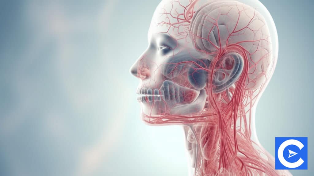
Carotid artery stenosis, myocardial infarctions, and ACS
Acute coronary syndrome (ACS)
This refers to impaired blood flow through the coronary arteries.
The result will be ischemia of the heart muscle, which can cause angina in severe cases.
Females, elderly patients, and diabetics can experience less acute symptoms.
Shortness of breath, numbness, and pain in the arms are common symptoms.
You should be aware of three types of angina classifications:
- Stable angina
- Unstable angina
- Variant angina
Myocardial infarctions
Non-ST-segment elevation MI (NSTEMI)
When myocardial ischemia or infarction causes severe myocardial damage, an ECG shows ST elevation.
If this elevation is not present, this could indicate NSTEMI or unstable angina.
Heart enzyme levels will increase if there are indications of partial blockages in coronary arteries when a NSTEMI is present.
The symptoms of unstable angina include tightness or pain in the chest that moves into the arms, anxiety, dyspnea, dizziness, weakness, nausea, vomiting, and heartburn.
Initially, beta-blockers, nitroglycerin, antiplatelet agents, and antithrombotic agents are prescribed.
ST-segment elevation MI (STEMI)
This sees ST elevation as a result of a complete blockage on an artery that has myocardial damage and is much more severe.
Symptoms related to this are the same as acute MI.
Necrosis may lead to irreversible myocardial damage, so immediate reperfusion is necessary to prevent death.
Myocardial infarction manifestations
Clinical manifestations of myocardial infarction are highly variable, and in more than 50% of patients that have cardiovascular disease, no prior history is present.
These are some of the symptoms:
- Angina
- Hypertension
- Palpitations
- Dyspnea
- Dependent edema
- Pulmonary edema
- Vomiting
- Nausea
- Pallor
- Cold and clammy skin
- Diaphoresis
- Lower urine output
- Disturbance, both neurological and psychological
- Headaches
- Slurred speech
- Lightheadedness
- Fear
Under this section in your coursework, you will find information regarding the symptoms and diagnosis of papillary muscle rupture and carotid artery stenosis.
You can catch up on them there, as we will not be going into any great detail about them in this guide.
Endocarditis, heart failure, and hypertensive crises

Heart failure
Heart failure is often characterized by symptoms such as pulmonary edema, peripheral edema, or systemic edema, along with contraction or filling disorders.
There are several causes of this condition, including systemic or pulmonary hypertension, coronary artery disease, valvular disorders, and cardiomyopathy.
Heart failure may be systolic or diastolic.
Classifying heart failure based on symptoms and prognosis is done from class 1 to 4, with IV having the worst prognosis.
Heart failure can be treated in various ways, including:
- Monitoring fluid balance and weight changes to determine changes in fluid retention
- Limiting sodium intake in your diet
- Patient activities restricted
- The use of medications to reduce the workload of the heart (such as vasodilators, ACE inhibitors, and diuretics)
- The use of anticoagulants
Although we won’t go into detail here, your coursework will cover systolic and diastolic heart failure as well as acute heart failure.
Know the symptoms, diagnoses, and overall treatment of these conditions.
Acute cardiac-related pulmonary edema
When heart failure occurs, fluid overload causes acute cardiac-related pulmonary edema.
A large amount of this fluid is then absorbed by the lungs, particularly in the interstitial spaces.
Pulmonary edema is caused by MI, chronic HF, mitral stenosis, ischemia, and volume overload.
Symptoms include:
- Coughing with sputum that includes blood
- On auscultation, there are crackles and wheezing
- Diaphoresis
- Severe dyspnea
Echocardiograms, x-rays, and auscultation can all be used to diagnose this, and there are many treatment options possible.
Hypertensive crises
As a result of the extremely high blood pressure, severe organ damage can occur if untreated.
This can happen due to eclampsia, noncompliance with prescribed medication, pulmonary edema, stroke, subarachnoid hemorrhage, or an aortic aneurysm.
This is characterized by anxiety, ruddy skin, sweating, headaches, blurred vision, and basilar HA, among other symptoms.
In addition to CBCs, BMPs, urine tests, ECGs, and chest x-rays, hypertensive crises can be diagnosed with blood tests.
Treatments include medication as well as intervention by medical staff.
Endocarditis
Infection of the heart valve lining causes this condition.
A number of risk factors contribute to this disease, including age (over 60), gender (mostly affects men), dental infections, and IV drug use.
In some cases, endocarditis can be treated with medication, but in others, it needs surgical repair of the valves.
Some serious complications of endocarditis include heart failure, sepsis, and emboli.
Diagnostic methods include:
- Blood cultures
- Echocardiogram
- ECG
- Anemia
- If the patient has a higher-than-normal count of white blood cells
- Elevated C-reactive protein
- Elevated erythrocyte sedimentation rate (ESR)
VTE, stenosis, and myocarditis

Myocarditis
Myocarditis is an inflammation of the heart muscle tissue or cardiac myocardium.
Most often, this occurs as a result of them having a viral infection, such as influenza or Coxsackie.
Myocarditis can also be caused by an allergic reaction to a medication the patient was prescribed, or by bacteria, fungi, or parasites.
There are some cases in which antibiotics and chemotherapy drugs can cause myocarditis
In myocarditis, the coronary vessels are infiltrated by blood cells between muscle fibers, causing the heart to dilate and muscle tissue to degenerate.
The heart becomes weak and enlarged because it cannot pump blood efficiently and as a result, congestive heart failure can occur.
Myocarditis is characterized by the following symptoms:
- Dyspnea
- Chest pressure and discomfort
- Palpitations
- Fatigue
For diagnosis, the following can be used:
- Radiograph of the chest area
- Ecg
- Echocardiogram
- Cardiac biopsy
- Cardiac catheterization
- Viral cultures
- Viral titers
- Polymerase chain reaction
Treatment options include:
- Treating the underlying cause through antibiotics
- Limiting activities
- Heart failure monitoring and treatment if necessary
- Oxygen
- In acute cases, gamma globulin can be administered through IV
Acute pericarditis
A patient with pericarditis has an inflamed pericardial sac with or without pericardial fluid accumulation.
In some cases, this can be a result of an underlying disease, but it can also result from a single event.
When the cause is malignant or autoimmune in nature, the patient will suffer from symptoms related to the cause.
In most cases, however, cases are caused by viral etiologies, which means patients have similar symptoms to people with influenza.
These are some of the symptoms and signs:
- Sharp chest pain. The symptoms get worse with inspiration, while leaning forward or sitting up alleviates them
- Pericardial effusion
- Respiratory distress
- Auscultated friction rub
- Pericardial effusion risk
- PR depression/ST elevation
Pericardiocentesis and pericardial biopsy, echocardiograms, ECGs, and elevations of WBC, ESR, and CRP can help provide a diagnosis.
In extreme cases, pericardiectomy surgery may be necessary but more often than not, it can be treated with medication.
Mitral stenosis
Flow of blood from left atrium to left ventricle is controlled by the mitral valve.
Mitral stenosis occurs when the valve narrows.
As a result, the left atrium gets bigger and pulmonary hypertension occurs since the pressure increases in the pulmonary veins and capillaries to overcome the resistance.
Infectious endocarditis as well as tumors in the left atrium may cause mitral stenosis.
There are several symptoms and signs of right side heart failure, such as loud S1 and S2 sounds, a mid-diastolic murmur, and dyspnea (exertional, orthopnea, and nocturnal).
In order to diagnose mitral stenosis, ECGs, echocardiograms, chest x-rays, and cardiac catheterization can be used.
The condition can be treated with medications, but in severe cases, surgery may be necessary.
You should also check your coursework regarding mitral valve insufficiency while on the subject of the mitral valve.
Aortic stenosis
In this condition, the left ventricle’s aortic valve narrows.
To overcome resistance from the valve, the left ventricle wall thickens, resulting in increased pressure.
Besides increasing afterload, this also increases coronary artery blood supply.
The development of aortic stenosis can be caused by rheumatic fever during childhood.
Symptoms include:
- Angina
- Intolerance to exercise
- Dyspnea
- Split S1 and S2
- At the base of the carotids, there will be a systolic murmur
- Exertion hypotension
- Syncope
- Failure of the left side of the heart
An x-ray of the chest, an echocardiogram, an ECG, and a cardiac catheterization can be used to diagnose the problem.
The condition might require surgery in severe cases, but it can usually be treated with medications.
Also in your coursework, you will find information on pulmonic stenosis that you should work your way through.
Blunt and penetrating cardiac trauma
Those who have been in vehicular accidents, fallen, or been hit on the chest in some other manner can suffer blunt cardiac trauma.
Their respiratory system may be affected, and their greater vessels might rupture as a result, leading to respiratory distress.
The intrathoracic pressure may also increase and cardiac tamponade may occur.
Due to their anterior location, the right ventricle and atrium are most likely to be injured.
A patient with suspected blunt cardiac trauma should receive an ECG as soon as possible.
If any abnormalities are found, continuous cardiac monitoring is recommended for the following 24 to 48 hours.
In order to diagnose, a CT scan or chest x-ray can be performed, as well as a transesophageal echocardiogram for verification.
To prevent a rupture, surgical repair may be required while internal injuries or clotting should be checked in vessels.
In cases of gunshot wounds or stabbings, the heart suffers penetrating cardiac injuries.
The mortality rate for these types of injuries is high within one hour of receiving them.
Instead of stabilizing patients first, they should be taken to a medical facility as soon as possible.
It is also recommended that you do not remove the object or object part causing the wound unless a physician is present.
These primary complications can arise from penetrating cardiac injuries.
In the case of a gunshot wound, you would usually experience exsanguination.
The patient may suffer hemorrhagic shock and hemothorax shock in this case, which have very poor prognosis.
It is more common to develop cardiac tamponade as a result of knife wounds.
Patient prognosis is good with surgical repair, however.
There are three common symptoms of cardiac tamponade, including muffled heart sounds, low arterial blood pressure, and jugular vein distention.
A triad of these three is known as Beck’s triad.
There is also pneumothorax, which is characterized by anxiety, tachypnea, rising SVR, and a deviated trachea.
There is an urgent need to treat this medical emergency.
Adulthood congenital heart defects
It is common for congenital heart defects to be picked up in early childhood, but sometimes they aren’t diagnosed until adulthood.
In addition, some can occur at any age, such as atrial and ventricular septal defects.
A congenital heart defect may present with the following symptoms:
- Shortness of breath
- Fatigue
- Syncope
- Palpitation
- Edema
- Murmurs
- Cyanosis
- Clubbing of the fingernails
Diagnosis can be carried out through a physical assessment, x-ray, EKG, echocardiogram, MRI, and CT scan.
A defect’s size and location determine its treatment.
The most common treatment is surgical repair.
Peripheral arterial and venous insufficiency
Peripheral arterial and venous insufficiency is characterized by these characteristics.
Arterial insufficiency
- Pain: Extends from intermittent claudication up to constant shooting pains that are severe
- Pulses: Absent or weak
- Skin: The skin is cool, pale, and shiny. Rigid and thick nails, while hairs on the toes and foot will disappear
- Ulcers: These will occur on the tips of the toes, heels, or other areas of pressure. They are extremely painful, circular in shape, and deep
- Edema: Minimal
Venous insufficiency
- Pain: Cramping/aching
- Pulses: Present/strong
- Skin: Ankles and anterior tibial area shows discoloration (brownish in color)
- Ulcers: Ulcers can be found on the medial or lateral malleolus and occasionally on the anterior tibial area. Ulcers are irregular and superficial with varying degrees of pain
- Edema: Moderate to severe
Learn more about acute peripheral vascular insufficiency in your coursework.
Cardio interventions and procedures

Cardioversion
A patient is sedated or anesthetized and a timed electrical stimulation is sent to the heart.
A normal sinus rhythm is then achieved by converting atrial fibrillation or other tachydysrhythmias.
As for how the procedure is carried out in full, that is already covered in your coursework.
Emergency defibrillation
In defibrillation, a patient receives a non-synchronized shock via pads or paddles at any time during the cardiac cycle.
There are several problems that can be treated with defibrillation, including acute ventricular fibrillation, pulseless ventricular tachycardia, and polymorphic ventricular tachycardia.
In order for a sinus rhythm to be regained, myocardial cells must be depolarized and then repolarized.
Your coursework should explain how defibrillation works.
Pericardiocentesis
The procedure can help diagnose pericardial effusion when performed under ultrasound guidance.
An ECG and ultrasound guidance can also be used to relieve cardiac tamponade.
Additionally, the procedure can be used to treat cardiac arrhythmias and PEAs accompanied by elevated jugular venous pressure.
It is possible to alleviate a non-hemorrhagic tamponade in 60 to 90% of cases, but hemorrhagic tamponade requires a thoracotomy in about 10% of cases.
Your coursework covers the procedure for a pericardiocentesis in detail.
Pacemakers
It is possible to implant a pacemaker when the heart’s normal conduction system is defective.
They can be permanent or temporary and will help stimulate the heart.
Pacemakers are used in the following clinical settings:
- To treat dysrhythmias that do not respond to medication and remain persistent
- To increase overall cardiac output by increasing the rate with bradyarrhythmia
- To lower ventricular/supraventricular tachycardia
- To treat secondary heart block
- After cardiac surgery, to improve cardiac output
- To provide diagnostic information
Transcutaneous pacing
When medication hasn’t helped with symptomatic bradydysrhythmias and hemodynamic instability has resulted, this type of pacing can be used as a stop gap.
Epicardial pacing
The epicardial pacing wires can be placed directly on the exterior atria, ventricles, or both following CBP or valve repair surgery when pacing support is required.
Temporary transvenous pacemakers
Patients with symptomatic bradycardias or heart blocks may benefit from these pacemakers, which consist of a lead at the end of a catheter.
As temporary treatments, they can either be used therapeutically or prophylactically.
In your coursework, you can read about how to set up all three.
Complications: Pacemakers
These include:
- Bleeding, infections or hematomas
- Hemothorax, caused by the internal mammary artery or the subclavian vein being punctured
- Ectopic beats or tachycardia may result from the ventricular wall being irritated by the endocardial electrode
- The myocardium may be perforated or there might be a pacemaker malfunction if the transvenous lead is dislodged
- During temporary pacing when the epicardial wires are removed, this can result in cardiac tamponade
- Phrenic nerve or muscle stimulation can occur as a result of dislocation of the leads
- Pacemaker syndrome
The last point, pacemaker syndrome, deserves a brief mention.
This can happen when the ventricles and atria do not contract in sync.
As a result, two things happen.
First of all, cardiac output is decreased.
Second of all, the atria are not sufficiently involved in filling the ventricles.
In addition to hypotension after decompensation, total peripheral vascular resistance rises to maintain blood pressure.
There are three categories of pacemaker syndrome: mild, moderate, and severe and in some cases, heart failure may result.
Automatic ICD
As with pacemakers, an automatic implantable cardioverter-defibrillator (AICD) is implanted in a patient in much the same way.
Fibrillation and/or tachycardia are controlled using this device.
The majority of modern devices can act both as pacemakers and ICDs, so they can be used on patients with bradycardia and tachycardia.
Closure devices with PCI
Percutaneous catheter intervention (PCI) is a treatment choice for patients with symptomatic coronary artery disease
This includes:
- Percutaneous transluminal coronary angioplasty (PTCA)
- Atherectomy
- Stent insertions
These will not be covered here, but you will need to read up on them in your coursework.
We should briefly discuss the potential complications associated with these procedures.
They include:
- Coronary artery spasms
- Dissection of that artery
- Bleeding
- Thrombosis
- Formation of hematomas
Coronary artery bypass graft (CABG)
The following conditions can be treated with this surgical intervention:
- Angina
- Unstable angina
- When the left main coronary artery is more than 60% blocked
- Multiple artery blockages
- Left ventricular dysfunction
- PCIs that were unsuccessful
There can be a variety of complications, including:
- Infections
- Arrhythmias
- Cardiac tamponade
- Cardiogenic shock
- Stroke/embolus
- Pulmonary or renal dysfunction
Learn about two types of CABG in your coursework: Minimally-invasive direct coronary artery bypass (MIDCAB) and Port access coronary artery bypass graft.
Cardiac valve repair: Surgical options
Heart valves can be repaired in a variety of ways:
- Valvotomy/valvuloplasty: The procedure involves opening stenosed valves or releasing adhesions between valve leaflets
- Aortic valve replacement: Aortic valves cannot be repaired, so they must be replaced
- Aortic homograft: A donor’s aorta and aortic valve are used to replace the recipient’s aortic valve and ascending aorta
- Ross procedure: A pulmonary artery and pulmonary valve are used to replace the aortic valve and a portion of the aorta. It is then replaced by a donor graft.
Cardiac surgery postoperative care
First, let’s examine the recovery period:
- It is important to monitor the patient’s hemodynamics closely, as well as to check their vitals every five to fifteen minutes
- While warming the patient according to procedures, the ECG should be assessed for arrhythmias
- It is recommended to maintain the MAP at 70-110 mmHg. This prevents graft hemorrhage and rupture, and inadequate perfusion. It should be possible for the patient to produce 30 mL/hr of urine
- Be sure that the chest tubes are not producing excessive amounts of fluid (less than 200 mL per hour) and that they are not decreasing or stopping altogether
- Auscultate heart sounds with the extremities elevated. Perfusion can be assessed this way
- Monitoring ABGS should be done every two to four hours during recovery. If necessary, adjust the ventilator settings
- Monitor blood sugar levels closely (if using insulin drips) and ensure cephalosporin prophylactically to prevent infection
- If necessary, replace electrolytes and monitor CMP
It is possible patients experience complications such as:
- Cardiac tamponade
- Hypotension/shock
- Arrhythmias
- Stroke
Insertion of an arterial line
Arterial lines are indicated as follows:
- Hemodynamic instability
- Regular ABG monitoring
- Placement of IABP
- Arterial pressure monitoring
- Administering medication when venous access is not possible
Sterile arterial line insertion can be performed at the following sites:
- Radial (which is most often used)
- Femoral
- Brachial
- Dorsalis pedis arteries
Your coursework should explain how an arterial line is inserted.
Complications may include:
- Infection
- Coagulopathy
- Bleeding
- Thrombosis
- Advanced atherosclerosis
Central line insertion
It is necessary to do this when administering large volumes of liquid to patients, such as for blood testing and CVP measurements.
Lines can be placed in a variety of locations, including the internal jugular vein, subclavian vein, and femoral vein (although this is not done frequently).
To locate the insertion site, an ultrasound will be used and then verified before insertion begins.
To find out how to install a central line, check your coursework.
Vascular interventions
During ischemia-related damage, the limbs of the patient will need vascular intervention when there is decreasing blood flow.
There are several reasons why vascular interventions could be necessary:
- Acute occlusion (embolus)
- Vascular disease that doesn’t respond to other treatments
- Dissecting/rupture aneurysm
- Damaged vessels
- Congenital defect
Among these interventions are:
- Bypass grafts
- Embolectomy
- Aortic aneurysm repair
Through careful monitoring and controlling of the patient’s blood pressure, the patency and integrity of the grafts are maintained.
As emboli may block cerebral or renal arteries, kidney and neurological function must also be monitored.
It is possible to experience complications following a graft transplant, including infection at the site of the transplant, pulmonary infection, kidney dysfunction, blood clots, hemorrhage, and occlusion.
Respiratory failure, ALI, and acute pulmonary embolism
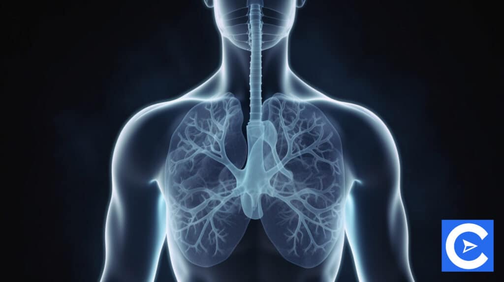
In the event of a blocked pulmonary artery or arteriole, pulmonary embolism could result.
There are several ways in which they can develop, but mostly as a result of thrombus formation.
Septic embolism, fat, or air can cause this condition.
Many thrombus formations originate in deep veins in the legs, right atriums, and pelvic veins.
Blood pools in the right atrium during atrial fibrillation, making this a very serious condition.
It is possible for them to enter directly into the lungs through the right ventricle.
An embolus can cause the following symptoms depending on the area of occlusion and its overall size.
- Dyspnea with tachypnea
- Cyanosis
- Anxiety
- Chest pain which can move onto arrhythmias
- Fever
- Rales
- Cough
- Hemodynamic instability
Diagnostic tests include:
- ABG analysis. This could indicate hypoxemia, hypocarbia, and respiratory alkalosis
- D-dimer: This can reflect as elevated PE
- ECG: This may show abnormalities such as sinus tachycardia
- Echocardiogram which may detect central artery emboli
- Spiral CT: This could give a definitive diagnosis
- V/Q scintigraphy will confirm diagnosis
- Pulmonary angiograms will confirm diagnosis
The following can be included in the medical management of this condition:
- Oxygen
- Intravenous infusions
- Various medications (antiarrhythmics, diuretics, digitalis glycoside)
- Cardiac monitoring
- Anticoagulants
- Analgesics
- Percutaneous venous filter placement
- Thrombolytic therapy
Acute respiratory distress syndrome (ARDS) and acute lung injuries (ALI)
ALI usually leads to ARDS, an extremely dangerous condition that can lead to death (5-30% mortality rate).
The following symptoms are present:
- Crackling rales/lungs wheezing
- Refractory hypoxemia
- Increased tachypnea with expiratory grunting because of a decrease in pulmonary compliance
- Skin mottling/cyanosis
- Tachycardia and hypotension
- Volume overload symptoms are missing (JVD or 3rd heart sound)
- Initially, the patient will have respiratory alkalosis. Hypercarbia and respiratory acidosis will replace this as the disease progresses
There are several treatment options available:
- To maintain ventilation and oxygenation, the patient can be put on mechanical ventilation
- Anti-inflammatory medication, inhaled surfactant, nitrous oxide, and corticosteroids
- A treatment plan for the underlying condition
- To reduce the number of days on a ventilator, conservative fluid management is necessary
Acute respiratory failure
Cardinal signs include:
- Tachypnea
- Tachycardia
- Anxiety, restlessness
- Diaphoresis
The following symptoms can occur depending on the cause:
- Depth and pattern of respirations changing
- There may be signs of cyanosis
- Central nervous depression and changes in overall consciousness
- Cardiac arrhythmias may occur
- With depressed respirations, dyspnea becomes more pronounced
- The patient falls into a stupor, coma, or could even die if there is no reversal in the condition
Make sure you examine both hypoxic and hypercapnic respiratory failures in your coursework.
In addition, let’s briefly discuss the underlying causes of respiratory failure:
- Airway obstruction
- Inadequate respirations
- Neuromuscular disorders
- Pulmonary abnormalities
- Chest wall abnormalities
Patients experiencing respiratory failure can benefit from the following nursing interventions:
- Place the patient in an ambulatory position by turning them
- Ensure that the patient coughs and breathes deeply
- Percussion and vibration treatments
- Make sure they stay hydrated. It helps keep airway secretions hydrated and encourages spirometry
Emphysema, bronchitis, and pneumonia
Pneumonia
Inflammation of the lung parenchyma and filling of the alveoli are signs of pneumonia.
There is a possibility that pneumonia may accompany another disease like lung cancer, as a secondary infection, or it may occur on its own, as a primary infection.
A variety of causes can lead to pneumonia, including fungi, parasites, viruses, and bacteria.
Hospital-acquired pneumonia occurs when a patient becomes ill with pneumonia while being treated for some other ailment in a medical facility.
Patients who contract pneumonia after being hospitalized in an acute care facility within 90 days are said to have healthcare-associated pneumonia.
In order to treat pneumonia, the following steps are taken:
- Antibiotics
- Pneumonia patients should be isolated and precautions taken
- It is important to ventilate patients in a 30-degree upright position, while ventilatory circuits should also be changed according to procedure for vent patients.
As part of your coursework, make sure you cover appreciation pneumonitis/pneumonia since we won’t go into detail here.
There are three components to look through: diagnosis, treatment, and symptoms.
Bronchitis
As a characteristic of this respiratory disease, a patient must cough and produce sputum for a minimum of three months each year for two consecutive years.
It is not uncommon for pollutants or smoke to irritate the airway, resulting in an inflammatory response.
As mucus-secreting glands produce more mucus, the airways are plugged, and ciliary function is reduced.
Additionally, the alveoli near inflamed bronchioles become fibrotic and bronchial walls thicken.
A compromised alveolar macrophage function means a greater chance of infection.
Chronic bronchitis symptoms include:
- Increasing sputum and a persistent cough
- Dyspnea
- Respiratory infections that occur often
The following treatment options are available for chronic bronchitis:
- Bronchodilators
- Oxygen therapy over a long term or during exercise, the use of supplemental oxygen
- Exercise and breathing can be improved with pulmonary rehabilitation
- It is possible to treat infections with antibiotics when they occur
- The use of corticosteroids is necessary to treat acute conditions
Make sure to review your coursework and the section on emphysema, of which there are two primary types: centrilobular and panlobular.
Chronic obstructive pulmonary disease (COPD)

COPD stages can be identified using functional dyspnea, BMI, and spirometry.
In spirometry, forced expiratory volume is measured for the first second of expiration (FEV1).
It is measured after full inhalation based on total forced vital capacity (FVC).
In total, there are four stages:
- Stage 1 which is mild
- Stage 2 which is moderate
- Stage 3 which is severe
- Stage 4 which is very severe
Despite COPD not being reversible, the goal is to slow it down and ease symptoms in order to improve overall quality of life.
In order to manage properly, you must:
- Prescribing necessary medications (if the patient smokes) to help them quit
- The use of bronchodilators
- The use of corticosteroids
- Oxygen therapy
- Bullectomy
- Lung transplant
- Pulmonary rehabilitation
Chronic asthma
There are three key symptoms to look out for in chronic asthma patients.
These are dyspnea, wheezing, and coughing.
The symptoms of chronic asthma include recurrent bronchospasms and airway inflammation.
The bronchi are the tissues that asthma affects, not the alveoli.
COPD does not include chronic asthma because it is not characterized by constant obstruction of the airway.
Symptoms of chronic asthma include:
- Exertional dyspnea
- Chest tightness
- Coughing at night
- General cough
The following treatments are available:
- Avoiding triggers
- Bronchodilators
- Chest hygiene
- Treating infection promptly
- Use of inhaled glucocorticoids
A severe acute asthma attack unresponsive to traditional treatments is known as status asthmaticus.
An attack of this type usually requires a trigger or stimulus.
A person afflicted with status asthmaticus will be in severe distress and will exhibit the following symptoms:
- Signs that their airway is obstructed
- Intercostal and sternal retractions
- Dyspnea and tachypnea with growing cyanosis
- Prolonged expirations that are forced
- Increased pulmonary edema
- Afterload increase in the left ventricle in cardiac decompensation
- Pulsus paradoxus
- Hypoxemia
- Hypocapnia and then hypercapnia
- Metabolic acidosis
Non-invasive ventilation and airway equipment
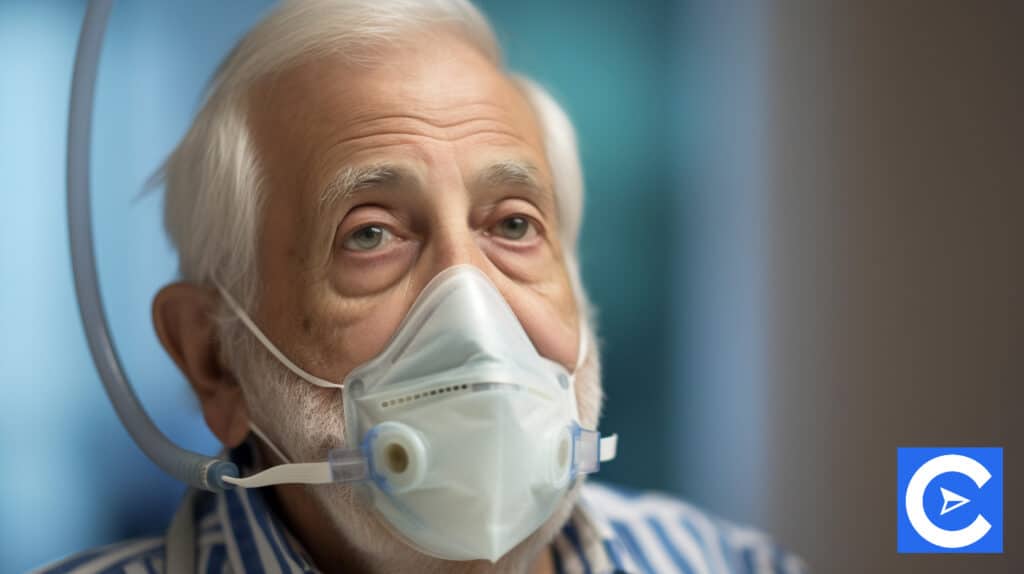
Non-invasive ventilation
There are quite a few pieces of medical equipment that will fall under this category that is thoroughly covered in your coursework and are used regularly.
These include:
- Nasal cannula
- Non-rebreather masks
- Non-invasive pressure ventilators (CPAP and Bi-PAP)
- High-flow and low-flow oxygen delivery devices
Airway devices
As for airway devices, these include the following tubes:
- Oropharyngeal
- Nasopharyngeal
- Tracheostomy
Chest tubes and mechanical ventilation
Mechanical ventilation
Endotracheal intubation
For patients with respiratory failure who suffer from hypoxemia, hypercapnia, hypoventilation, or who have an obstructed airway, endotracheal intubation is often required.
Ensure you cover how this is done in your coursework.
The following methods can be used to place endotracheal tubes correctly:
- Capnometry
- Capnography
- Esophageal detection devices
- Chest x-rays
Management of the ventilator
There are many types of ventilators in medical facilities, but the basic principles of ventilator management are the same.
As a result, it is necessary to monitor the following:
- Ventilation type
- Control mode
- Tidal volume
- Inspiratory-expiratory ratio
- Respiratory rate
- Fraction of inspired oxygen
- Sensitivity
- Pressure
- Rate of flow
A patient who suffers a ventilation-induced lung injury is said to have suffered a VILI or ventilation-induced lung injury,
In patients with ARDS, however, this is common.
VILI is made up of four interrelated components:
- Barotrauma: Excessive pressure causing damage to the lung
- Volutrauma: High tidal volume causing alveolar damage
- Atelectotrauma: The repeated opening and closing of alveoli results in injury
- Biotrauma: This is an inflammatory response
Preventing ventilator complications
These include:
- When the patient’s head and chest are elevated to 30 degrees, aspiration and ventilator-associated pneumonia can be prevented
- Repositioning should be done every two hours for the patient
- It is recommended that the patient receive DVT prophylaxis
- Famotidine or pantoprazole PO/IV daily can prevent gastrointestinal ulcers and bleeding
- It is important to reduce and eliminate sedation/analgesia as soon as possible
- A barotrauma prevention protocol must be followed for pressure settings
- Keep an eye out for signs of barotrauma and pneumothorax
- Perform nutritional assessments and monitor for malnutrition
- Monitor intake and output to prevent dehydration
Check your coursework for information about ventilation weaning.
Failure to wean from ventilation
Twenty to thirty percent of patients fail to wean off of ventilation.
This can be affected by a variety of factors, including physical, situational, and psychological ones.
Taking a patient off ventilation requires them to protect their airways.
In addition, a solution must be found for the problem that led to ventilation initially as and there must be hemodynamic stability for the patient.
Weaning protocols are based on clinical criteria such as blood pressure, oxygen saturation, tidal volume, and respiratory rate.
Symptoms and signs of failure to wean from ventilation include:
- Lower tidal volume
- A respiratory rate that is increased
- Increased PaCO2
- Oxygen desaturation
- Fatigue
- Anxiety
- Diaphoresis
- Blood pressure and heart rate changes
- Hemodynamic changes
- Changes in mental state
Sedation with mechanical ventilation
Patients undergoing mechanical ventilation will initially receive sedation and/or analgesia.
This will be reduced over time as ventilation time increases with continuous patient sedation.
Sedation is used to:
- Ensure that movements or agitation do not interfere with ventilation
- A reduction in overall discomfort and pain
- Assist in controlling respiratory distress
Bronchoscopy
As part of performing a diagnosis, a flexible, thin fiber-optic bronchoscope is used to inspect the larynx, trachea, and bronchi.
Additionally, it can obtain biopsies, collect specimens, extract secretions or foreign bodies, excise lesions, and treat atelectasis.
Before this procedure, nares and oropharynx need to be anesthetized.
Chest tubes
Chest tubes are left in place on a patient who has undergone thoracic surgery or has suffered a pneumothorax as they will help drain any fluid or air, as necessary.
It will be necessary for these patients to receive adequate pain management.
In addition to suturing assistance, sterile techniques may also need to be observed, and dressings will need to be replaced on a regular basis.
Suction control, water seal, and a collection chamber comprise the test tube drainage system that’s in use.
Following chest tube placement, nursing interventions include reporting on drainage, checking the tube for occlusion after position changes, maintaining sterile dressings, checking for crepitus or drainage, and ensuring that the entire system is functioning properly.
Brain tumors, neuromuscular disorders, and seizures

Multiple sclerosis
It is an autoimmune disorder that affects the central nervous system.
In this case, the myelin sheath surrounding nerves is damaged.
As a result, nerve pulses can no longer be conducted because of scar tissue which forms.
It can cause tremors, speech slurring, vision impairment, pain, bladder and bowel dysfunction, and balance and coordination issues, among other symptoms.
An MRI and neurological and clinical examinations will be needed to diagnose the condition.
Treatment includes:
- Glucocorticoids
- Immunomodulators
- Immunosuppressant
- Hormones
ALS
There is a progressive degeneration of upper and lower motor neurons in this disease.
With time, symptoms such as hyperreflexia, muscle weakness, spasticity, and paralysis become more severe.
ALS is treated with:
- Antibiotics to control infection
- Mechanical ventilation
- Nebulizer treatments using steroids and bronchodilators
Other neuromuscular disorders discussed in your coursework (and which you can read about there) include Parkinson’s disease, Guillain-Barre Syndrome, and muscular dystrophy.
Seizure disorders
Generally, seizures can be divided into two types: partial and generalized seizures.
A partial seizure can be further classified into simple partial, special sensory, and complex partial.
Generalized seizures fall into two categories, tonic-clinic (Grand Mal) and absence (Petit Mal).
Epilepsy
There will be a history of seizures in patients suffering from this disease that is confirmed by EEG findings.
Each patient’s treatment is customized, but most will receive antiepileptic medications tailored to their unique needs.
It is always important to closely monitor patients taking certain epileptic drugs to avoid toxic reactions which can occur from time to time.
Brain tumors
It is possible to classify brain tumors as primary or secondary.
The origin of primary tumors is the brain, while metastasizing tumors occur in other parts of the body.
The following is a list of tumors you can learn about in your coursework:
- Astrocytoma
- Glioblastoma
- Brain stem glioma
- Craniopharyngioma
- Meningioma
- Ganglioglioma
- Medulloblastoma
- Oligodendroglioma
- Optical nerve glioma